At Eyecare Greengate we use cutting edge technology to identify, diagnose, and treat your conditions. Some new technology we are proud to offer below.

Optomap California
California icg was developed for retinal specialists to optimize management of AMD, uveitic conditions and other choroidal pathology. Imaging modalities and image viewing options are detailed below.
With California, Optos has incorporated new hardware and software technology enabling practitioners to see more, discover more and effectively treat more ocular pathology thus promoting patient health.

OPD Scan III wavefront aberrometer
Our OPD-Scan III Wavefront Aberrometer is an Autorefractor, Keratometer, Pupillometer (up to 9.5mm), Corneal Topographer, and Integrated Wavefront Aberrometer. The OPD-Scan III completes 20 diagnostic metrics in less than 10 seconds per eye (including angle kappa, HOAs, average pupil power, RMS value, and point spread function).
Easy alignment and automatic capture of wavefront aberrometry data ensures accurate readings. Wavefront aberrometry data is gathered from available zones up to a 9.5mm area, adding the capability to provide for the calculation of mesopic refractions. Blue light, 33 ring, placido disc topography is gathered in one second. Mapping methods include OPD, Visual Acuity Corneal Topography/Topographer, and more.
- Wavefront aberrometry with 2,520 light vector data points
- Corneal topographer/topography with 11,880 data point mapping
- Retro-illumination
- Day/Night Rx
- Auto X, Y, Z eye tracking
- Discerns AR vs. WF patients and their starting refraction point
- Sphere measurement -20D to +22D. Cyl 0.00D to ±12D
- Large, tilt LCD screen for superior viewing angle and operational position options

Corneal Topography
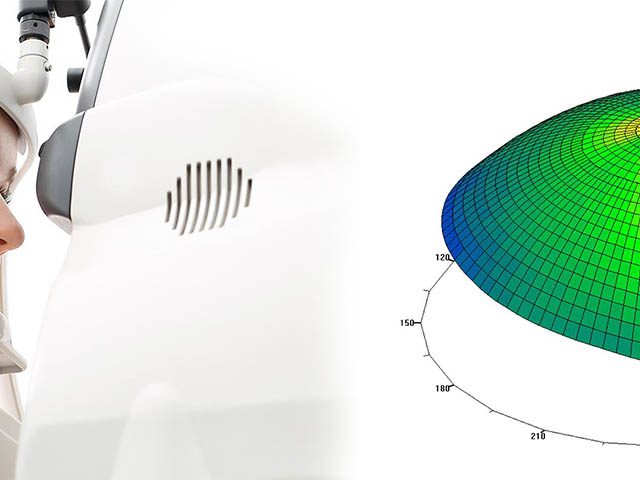
Your cornea is responsible for nearly 70% of your eye’s refractive power and therefore an important element in determining the quality of your vision. The Corneal Topography procedure available at Eyecare Greengate is non-invasive and used to map the curved surface of the cornea. During the test, you will be seated facing an illuminated pattern, which will be reflected on your cornea and will then reflect the back into a digital camera. The result of this technique is a three-dimensional map, which your doctor can examine in order to diagnose and treat a variety of conditions, plan for refractive (LASIK) surgery and help assist in fitting you with contact lenses.

Optical Coherence Tomography
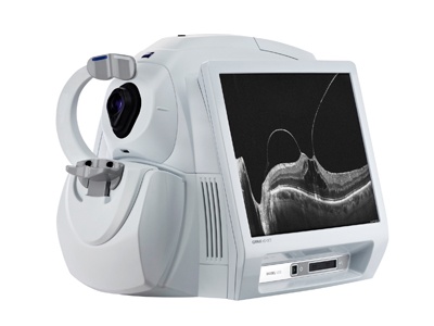
Eyecare Greengate is proud to offer Optical Coherance Tomography (OCT). OCT is used to image a precise area of the retina. OCT allows us to verify the health of your eyes by checking for certain abnormalities in the retinal structures, helping us diagnose conditions such as; macular holes, macular swelling, and glaucoma. OCT also allows us to monitor age-related macular degeneration and track how well you are responding to treatment. An OCT scan uses light beams, which are focused onto the back of the eye in order to produce detailed images of the inside of the eye. This test takes just a few minutes and you may need drops to dilate your pupils prior to testing.

Optical Coherence Tomography Angiography
Optical Coherence Tomography Angiography, or OCTA, is new, non-invasive approach to visualizing retinal vasculature that is transforming the way physicians see retinal and choroidal vasculature.
OCTA produces ultra-high resolution, three-dimensional images that are displayed as individual layers of retinal vasculature, allowing you to isolate specific areas of interest and see microvasculature that is not easily seen with FA or ICGA. New high-density OCTA imaging produces larger format scans with outstanding image quality to enable assessment with a wider field of view.

Humphrey Visual Field
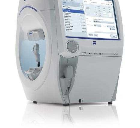
Developed by Carl Zeiss Meditec, Humphrey Visual Field (HVF) is revolutionizing the optical industry. HVF will depict a patient’s complete field of vision by drawing a “map” of it through the use of a simple process whereby a moving light or shape is projected in front of a patient and the patient is asked to indicate when it moves out of their peripheral vision. Your doctor may ask you to repeat the test in order to ensure the results are the same. In the case of glaucoma patients, this test is done once or twice a year to check for changes in vision. HVF are painless and quick – in most cases the test only takes three or four minutes.

Visioffice® System
Eyecare Greengate is the first practice in the Greensburg area to utilize the Visioffice system. Patented by Essilor, the leading lens manufacturer in the world, this revolutionary, state-of-the-art equipment provides eyecare professionals with a fast and accurate way to calculate measurements, ensuring you get the best fit possible with the most advanced lenses available. Visioffice is the first and only system that measures not just the physical contours of your eye – it also calculates how your eyes move. Taking into account your specific eye movements makes it possible to craft lenses that give you the most precise vision possible, no matter where you look through the lens.

Marco TRS 5100 Digital Automated Refraction System
The TRS 5100 benefits from Marco’s latest generation of electronic refraction technology. Replacing the standard refractor, it allows practitioners to control the entire refraction process from a keypad small enough to sit in your lap. This keypad also controls the CP-690 Automatic Chart Projector. And because the TRS 5100 is completely programmable, all the lenses are moved for you at the touch of a button, taking you to each new refraction step. While convenient for you, it is also helpful if you delegate refractions and want your technicians to perform the refraction steps in a specific order.

Meyefit Mirrors
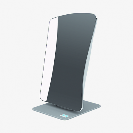
The m’eyeFit® mirror digital measuring system offers a reliable solution that delivers a modern patient experience. The plug-and-play designs make the device easy to set up for simple, fast, and accurate measurements.
Patient Benefits
- Quick, comfortable process
- Precise measurements for the latest lenses
- Personalized visual solutions for optimized vision
Eyecare Professional Benefits
- Simple and reliable measurement protocol
- Easy integration into current dispensing workflow
- Powerful processing capability
- Dedicated service and support
- Access to advanced personalized lenses in both single vision and progressive
Corneal Refractive Therapy
Corneal Refractive Therapy (CRT®) is a non-surgical process that gently reshapes your cornea while you sleep with the use of specially designed, gas-permeable contact lenses. CRT gives you clear vision during the day without the use of glasses or contact lenses. Because you wear CRT lenses only while you sleep, this option may actually be healthier for your eyes because it reduces lens wear time for most people. CRT changes are temporary; if you stop using the CRT night lenses, your vision will return to its original state in a day or two. If you have a low to moderate nearsightedness, with or without astigmatism, you might be a candidate for CRT. Ask our doctors here at Eyecare Greengate if you are a candidate for this exciting option!
Digital Retinal Photography
Digital Retinal Photography is the process by which a specialized low power microscope fitted with a digital camera takes a picture of your eye in order for your doctor to evaluate it for problems such as; diabetic retinopathy, glaucoma, macular degeneration, retinal detachment, hypertensive retinopathy and holes in the retina. Digital Retinal Photography, a non-invasive and painless process, is used to keep tabs on the health of your optic nerve, macula and the blood vessels of the retina. These pictures will become part of your patient record at Eyecare Greengate so that we can detect changes to these structures from year to year. Regular performance of Digital Retinal Photography is paramount to detecting problems early. At Eyecare Greengate, we believe early detection can make a huge difference in the treatment of your eye condition.
Anterior Segment Photography
Anterior Segment Photography involves the recording of the anterior structure of the eye, which includes the cornea, iris, ciliary body and lens. Eyecare Greengate completes this test to help document problems such as; cataracts, conjunctivitis, corneal ulcers or swelling. During this process a high-intensity light source, called a slit lamp, is used in conjunction with a biomicroscope for your doctor to fully examine the anterior segment.
